
A PhD student counts cells for 3 hours. The AI counts the same slides in 90 seconds, and it does not disagree with itself between Tuesday and Friday.
Cell counting is one of the most repetitive tasks in biology, pharmacology, and medical research. Every cell culture experiment, drug dosing study, and viability assay starts with the same question: how many cells are in this sample? The traditional answer involves a hemocytometer, a microscope, and a lot of patience. AI-powered counting tools now deliver results in seconds with better consistency than even experienced operators.
The manual counting grind
Manual cell counting with a hemocytometer follows a ritual that has barely changed in a century. Load the sample onto the counting chamber. Place the coverslip. Focus the microscope. Count four corner quadrants. Calculate the average. Multiply by the dilution factor. Record the number. Repeat for the next sample.
A single sample takes 10 to 15 minutes for a skilled technician. A typical cell culture day might involve 10 to 30 samples, and experiments that require time-course monitoring multiply that across hours or days. The math adds up fast: a research group running multiple assays can easily spend 20 to 30 hours per week on counting alone.
The bigger problem is not the time - it is the variability. Studies show that inter-operator variation in hemocytometer counting can reach as high as 52%, and even a single operator produces up to 20% variation between repeated counts of the same sample. Chamber loading errors contribute roughly 4.6%, pipetting adds another 4.7%, and even coverslip positioning introduces a 7.6% difference. When you stack these error sources, reaching better than 15% coefficient of variation requires counting hundreds of cells across multiple chambers.
How AI cell counting works
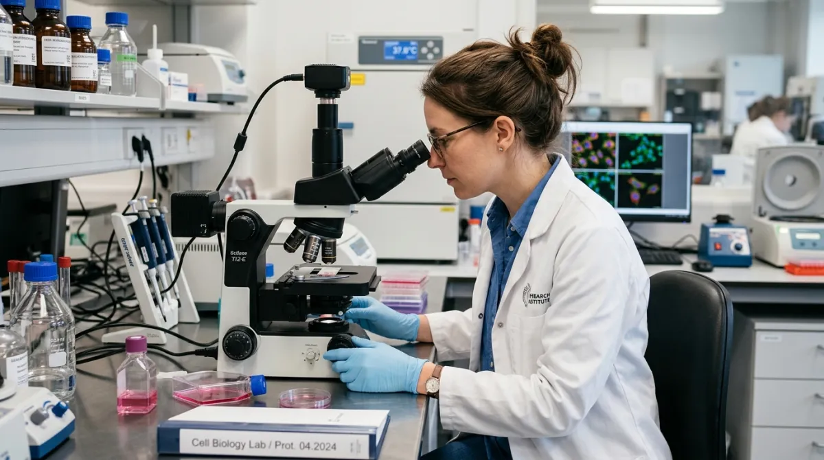
AI cell counting starts with the same input: a microscope image. The difference is what happens next. Instead of a human squinting at quadrants, a computer vision model segments the image, identifies individual cells, and returns a count with confidence markers - typically in under 30 seconds per image.
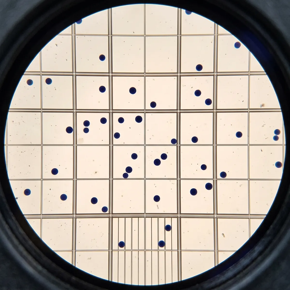
Tools like SnapCyte work directly with standard hemocytometer images captured at 10X magnification via a microscope camera or even a smartphone adapter. The AI detects the gridlines automatically, identifies cells within counting regions, and calculates concentration and viability. It supports Neubauer, Improved Neubauer, and Burker chamber types without manual configuration.
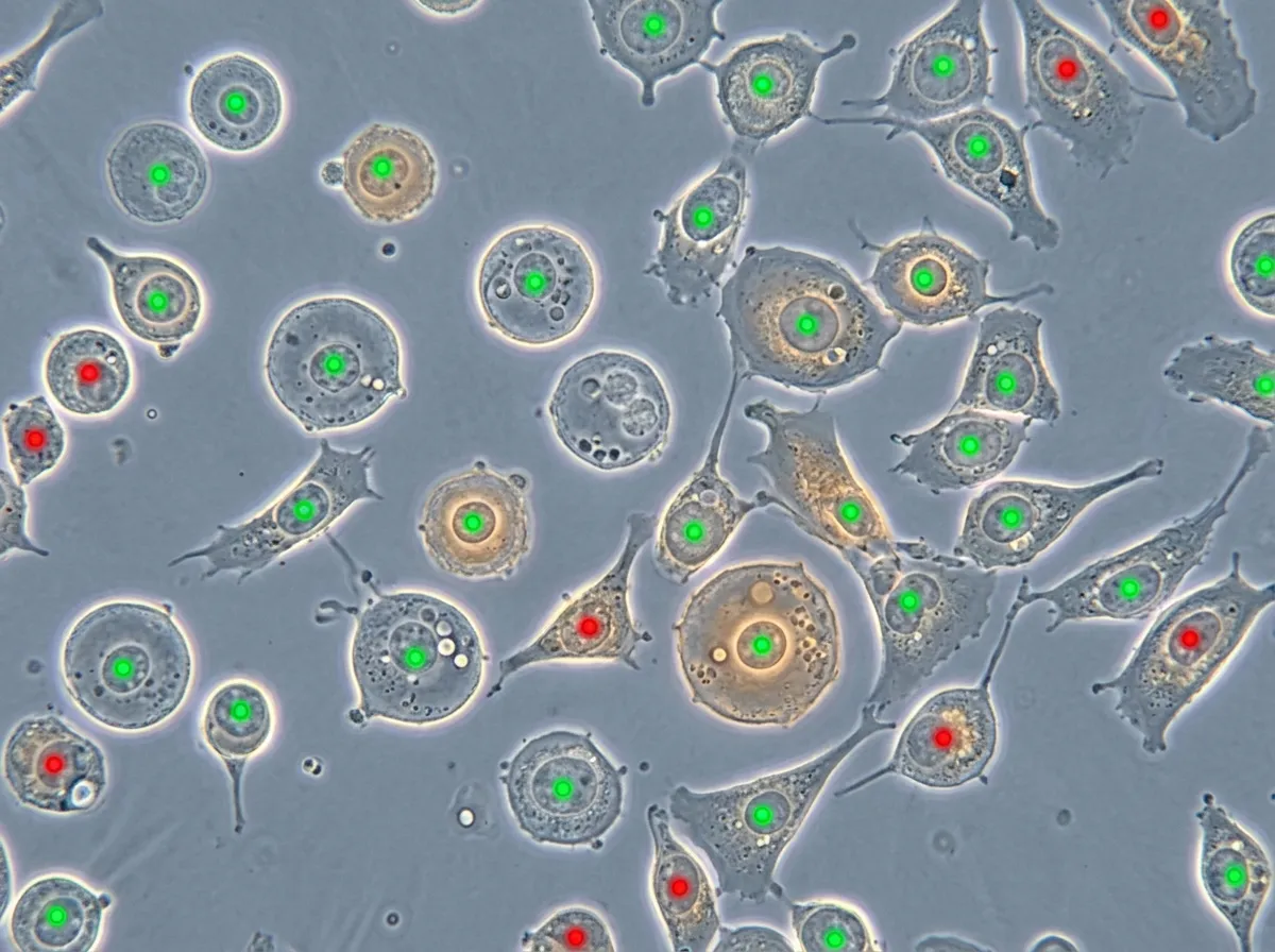
For adherent cells in culture plates, AI-enhanced microscopy uses phase-contrast or fluorescence imaging to detect cells directly in the vessel. No trypsinization, no sample transfer, no hemocytometer. The cells stay undisturbed in their growth environment.
Accuracy and reproducibility
AI cell counting achieves a mean absolute percentage error of less than 6.26% for live and dead cell mixtures - substantially better than the 15 to 52% variation range of manual hemocytometer counting. More importantly, the AI produces the same result every time it processes the same image.
Reproducibility is the real advantage. In drug screening, a 20% variation in baseline cell counts cascades through every downstream calculation: IC50 values shift, dose-response curves wobble, and experiments need more replicates to reach statistical significance. Consistent counting at the start tightens every result that follows.
10 to 15 minutes per sample. Coefficient of variation: 5 to 15% for experienced users, up to 52% inter-operator. Results depend on who counts and when.
Under 30 seconds per image. Near-zero inter-operator variation. Same image, same count, every time.
Cell types that work well
AI counting handles a broad range of cell types and preparations.
- Suspension cells (CHO, Jurkat, PBMC): straightforward detection with trypan blue viability staining
- Adherent cells in culture: phase-contrast imaging counts cells without lifting them from the plate
- Blood cells: white blood cell differentials and platelet counts from stained smears
- Bacterial colonies: count CFUs on agar plates from a single photograph
- Yeast cells: budding and non-budding differentiation for brewing and biotech applications
The main challenges are heavily overlapping cell clusters, dense debris fields, and very small cells below the resolution limit of the microscope objective. For most standard cell culture work at 10X to 20X magnification, AI counting is production-ready.
Getting started without new hardware
The barrier to entry is lower than most researchers expect. AI cell counting tools work with existing microscope setups - no specialized automated counter required. A standard lab microscope with a camera attachment is sufficient. Some tools even work with smartphone images captured through the eyepiece.
For labs already using hemocytometers, the transition is immediate: capture the image you would normally count by eye, upload it, and get a count with a confidence overlay. The hemocytometer stays, the squinting goes.
The bottom line
Cell counting has been a manual, subjective bottleneck in research labs for over a century. AI does not change the biology - it changes the time, consistency, and confidence of the count. Under 30 seconds instead of 15 minutes. Less than 6% error instead of 15 to 52% variability. The same result on Tuesday and Friday.
The next time a cell culture experiment begins with 30 minutes of hemocytometer counting, try photographing the chamber instead. The count will be ready before the incubator door closes.